
Eye exams have come a long way! If it’s been a while since your last eye exam, you may be surprised to see how far eye care technology has come.
Digital eye exams focus on using modern technologies for greater precision when screening for eye concerns and determining your vision prescription.
Whether you’re here to update your vision prescription or monitor your ocular health, a digital eye exam offers meaningful insights into the well-being of your eyes.
The Digital Eye Exam Defined
A digital eye exam refers to the incorporation of modern technology into the eye exam process. This contrasts with more traditional assessments that rely more heavily on physical tools and manual measurements.
Digital eye exams help us identify signs of eye diseases, monitor existing conditions, and even improve the accuracy of your prescription. It’s an all-encompassing process that treats your eye health with the care and precision it deserves.
As a general rule, we recommend:
- Children have annual exams until they turn 18 years old. They can have their first exam as early as 6 months!
- Adults (19-64) with no vision or underlying health concerns can get by with an eye exam every two years. But annual visits are recommended.
- Adults over the age of 65 should have annual eye exams as they are more likely to develop age-related visual changes.
The Technology Behind Digital Eye Exams
Here’s a breakdown of some of the instruments our expert team uses during your routine eye exam:
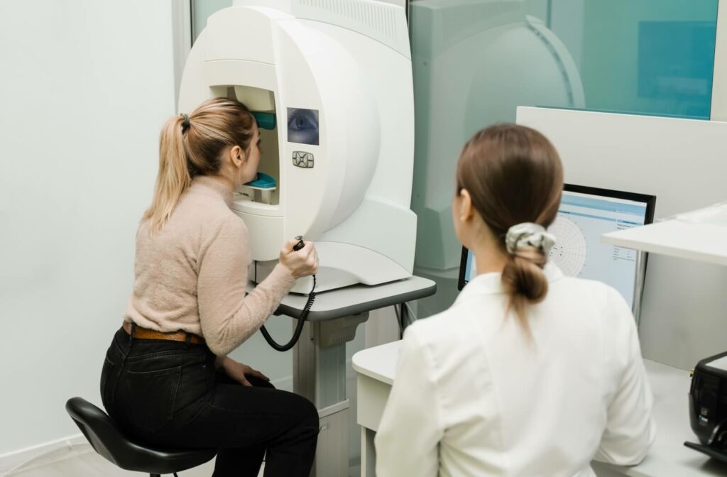
Optical Coherence Tomography (OCT)
OCT produces high-resolution, cross-sectional images of your retina. Simply rest your head against the device, focus on a single spot, and try to keep your eyes wide open. In a few short seconds, you’ll be able to see the layers of your retina!
OCT scans are instrumental in detecting fine changes in retinal layers that could signal macular degeneration, diabetic eye changes, or glaucoma. These scans offer an invaluable way to stay ahead of conditions that can significantly alter your vision.
Autorefractors
Say goodbye to the days of worrying about “Which is better, 1 or 2?” Remember, if you’re unsure, don’t hesitate to tell us!
Autorefractors provide an estimate of your current vision prescription. This machine generally requires you to focus on a single image (usually a hot air balloon or a barn) and measure how light travels through your eyes to determine an estimate of the lens power you need.
Autorefractors are also used for keratometry to measure the curvature of your cornea, which is helpful for your contact lens fittings.
We use these values during your refraction test to develop your finalized prescription. If you’re unsure about your lens choices during your eye exam, the measurements from the autorefractor can help us determine what might be a better fit.
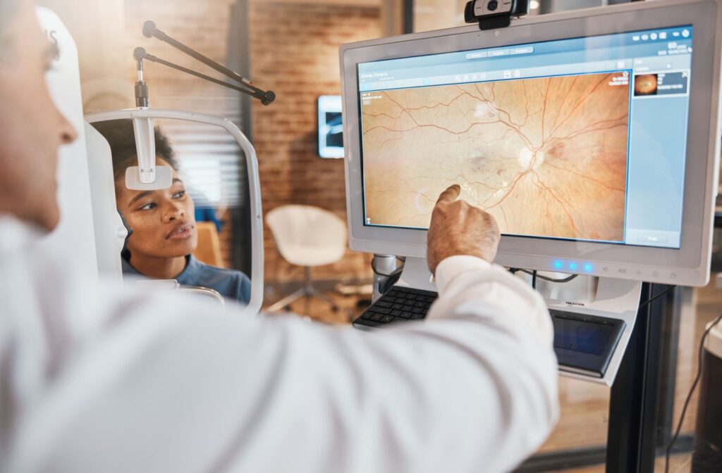
Fundus Photography
This instrument seamlessly complements OCT scans. Fundus photography captures detailed images of your retina, optic nerve, and blood vessels. These photos help us track changes in your eye health and identify early signs of various conditions.
Wait for the flash, keep your eyes wide open, and say cheese! In just a few seconds, you’ll be able to see what your eye’s internal structures really look like!
Ophthalmoscope And Slit Lamp
An ophthalmoscope is like a flashlight your optometrist uses to gain a closer look at the back of your eye. This helps your eye doctor spot warning signs of concerns like a retinal detachment or optic nerve damage.
The slit lamp works like a microscope for your eyes. Your optometrist uses this tool to inspect the front and back parts of your eyes in 3D.
A slit lamp exam can help identify concerns like cataract progression or severity, corneal damage, and signs of infections.
Visual Field Testing
Poor peripheral vision can be a warning sign for glaucoma. A visual field test is how we can assess a person’s peripheral vision.
A visual field test requires you to focus on a central target while small lights will appear across your field of vision, pressing a button whenever you notice them. The machine then maps out your responses, creating a detailed map of your visual field.
Keep in mind that most people may notice there’s a slight learning curve with this test.
Take Charge Of Your Eye Health
At Dr. Jennifer L. Shane & Associates, we carefully balance technology with health to deliver precise and trustworthy vision care.
Our digital eye exams allow us to look beyond the surface, offering detailed insights to empower you to make better decisions about your health. From early detection to updating your vision prescription, we’re here to help keep your eyes healthy and sharp. Connect with our team to schedule an appointment for your digital (routine) eye exam!